Fotofinder – Early diagnosis of melanoma
Skin cancer diagnostics in Katowice
FotoFinder is a professional and ultra-modern technology that enables analysis of the entire skin surface through automatic mapping and accurate diagnosis of individual nevus in just a few minutes. In addition, the FotoFinder Bodystudio ATBM II device, unlike a traditional videoscope or dermatoscopy examination, with its hi-tech solutions, allows for very precise documentation of the skin condition and individual nevus, thus enabling precise comparative analysis and early detection of cancerous changes on the skin. FotoFinder examination allows Patients to regularly monitor the appearance of the entire skin surface and lesions of different nature, which is the basis for prevention of skin cancer, including melanoma.
What is FotoFinder Bodystudio ATBM II?
The innovative FotoFinder Bodystudio ATBM II creates a map of the nevus that are on the body in just minutes. Using the Moleanalyzer pro AI Score software, the skin lesions are then immediately and accurately analyzed for health risks based on a developed algorithm. As the entire body surface is monitored during the examination, nevus located in areas that are difficult to inspect on their own are also analyzed. In addition, the FotoFinder Bodyscan software used in the device allows to compare images taken during the examination with those taken during follow-up visits. Thus, we are able to actively monitor the status of existing skin lesions and catch new ones that are potentially dangerous to the Patient’s health.
FotoFinder Bodystudio ATBM II examination, especially if performed regularly, allows for the detection of malignant lesions and melanoma at a very early stage. It is important to remember that skin cancer is 99% curable if diagnosed early enough and the appropriate treatment is implemented as soon as possible. FotoFinder allows for precise assessment of nevus, which is crucial in the diagnosis of dermatological diseases, including melanoma.
Indications for testing
- newly formed nevus
- mechanical and chemical skin irritations
- change in or around the nevus area (change in color, size, disappearance of the lesion)
- other troubling symptoms in or around the scar area such as oozing, excessive tenderness, etc.
- large number of birthmarks on the body
Additionally, the FotoFinder examination is also dedicated to individuals:
- frequent sunbathers and solarium users
- easily sunburned
- fair
- who have a family history of skin cancer
Course of examination:
The duration of the entire examination including analysis depends on the number of skin lesions of the Patient. Mapping the lesions with FotoFinder Bodystudio ATMB II takes only a few minutes. However, the examination can also include a more detailed analysis of individual nevus, in which case the time of the visit is longer.
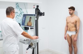
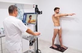
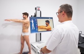
In our Clinic we use an innovative FotoFinder Bodystudio ATMB II platform, which enables monitoring of the whole skin surface and individual nevus thanks to ATBM® (Automatic Total Body Mapping) technology. During the examination, the Patient is asked to strip down to their underwear and stand in front of the camera in strictly defined positions. The physician then takes a series of photos of the entire body. In just a few minutes a map of the nevus on the Patient’s body is created. Using the Moleanalyzer pro AI Score software, the nevus photographed are thoroughly evaluated based on an algorithm developed over many years of clinical research. This algorithm classifies each nevus and detects those potentially dangerous to the Patient’s health. If historical Patient results exist in the database, the software performs a comparative analysis and captures any changes within pre-existing nevus and detects any newly formed ones. The mapping result is generated by the device in the form of a report for the physician and Patient. The physician then evaluates the result and makes a decision on whether to observe or treat the changes.
FotoFinder Bodystudio ATMB II also enables to perform videodermatoscopy with a special innovative head Medicam®. Videodermatoscopy consists in visualizing the examined skin fragment at high magnification on a computer screen. The physician performing the examination puts the camera to the examined skin area, including the scalp (using the TrichoLab module) or fingernails. The technology used in Medicam® allows the physician to obtain an accurate macro image in the required optical magnification with high HD resolution. Thanks to the image of very high quality and appropriate parameters the structure of the lesion is visualized, which is the basis for diagnosis, whether it has alarming features or not.


Technology
FotoFinder Bodystudio ATMB II examination is a versatile and effective examination due to the irreplaceable combination of high-tech equipment and special, innovative software. All this makes the FotoFinder examination significantly better than dermatoscopy or videodermatoscopy. FotoFinder is a highly developed, very precise and sensitive process for mapping and analyzing lesions on the entire skin surface, aiming at an early diagnosis of skin cancer.
The entire FotoFinder philosophy is based on the innovative ATBM® Automated Total Body Mapping procedure, which allows the entire skin surface to be analyzed and documented in a very short period of time and automatically capture potentially dangerous lesions at an early stage of their development.
ATBM® Automated Total Body Mapping or FotoFinder’s patented method of skin lesion mapping is based on very high accuracy, because diagnosis of nevus and comparative analysis of their condition can be effective if the examination is characterized by precision. ATBM® assumes:
- photographing and mapping the entire skin surface guaranteeing consistent documentation of the treatment areas. This is possible by fully automated camera positioning and the FotoFinder Ghost function, which helps position the Patient in front of the lens in a precise position determined by the laser light, making the maximum body surface visible, in exactly the same way during each examination.
- automatic diagnosis of all lesions on the Patient’s body. Moleanalyzer pro AI Score, an innovative solution based on the use of artificial intelligence, evaluates both melanocytic (pigmented) and non-melanocytic lesions (all other skin lesions) and, using the Deep Learning algorithm, determines whether a lesion is benign or potentially dangerous to the Patient’s health. This algorithm achieves impressively high results in terms of sensitivity and detail in evaluating skin lesions. In studies conducted, the algorithm has been shown to be so effective in diagnosing lesions that it can compete with experienced physicians in this regard. By constantly improving the algorithm and supplementing it with new data, image analysis becomes more precise.
- ability to make unambiguous comparative analysis of the same skin sections photographed at different points in time with the innovative Follow Up feature, which allows the images to be overlaid and automatically compared. Any changes that have occurred on the Patient’s body since the last examination are then captured and marked, so that none of them escape the physician’s attention and can be further diagnosed. This allows for effective control of the changes that have occurred and assessment of the possible progress of the disease. Precise comparison of images is possible thanks to:
- Standardized, repeatable images, which means that the Patient is always photographed according to a specially developed pattern, which involves taking a series of 20 images of the whole body skin from 4 sides in 8 standard positions.
- Documentation of nevus located throughout the body with all procedures in place to protect sensitive data. Maintaining a history of examination results allows to monitor the condition of skin and nevus over time.
- use of the highest quality camera and lens, allowing to create images and overview videos in very high resolution, which translates into precise diagnostics. The CrystalView technology used in the Full HD medicam® 1000s® camera with optical zoom allows for an even more precise "visual journey" into the skin, which enables the skin structures to be examined precisely at the cellular level. The visible lesion can be magnified up to 400 times and the image remains crystal-clear. The unique, built-in TwinLight irradiation allows to adjust the optimal light intensity to the lesion under examination and to operate with polarized and non-polarized light.
Report
It is worth mentioning that the Patient receives his/her copy of the result in the form of a report generated by FotoFinder. This provides the Patient with information on the analysis result, which allows for a better understanding of the diagnosis by the physician and decision-making process regarding possible follow-up or treatment. In addition, an additional copy of the report makes it easier for the Patient to perform regular self-examination of skin lesions on the body.
Repeating the test
It is recommended that the FotoFinder test be repeated once a year. For Patients with suspicious skin lesions, the physician may recommend repeating the examination every 30-90 days.
Experience is worth trusting – FotoFinder brand
FotoFinder is a German company with over 25 years of experience in developing skin imaging systems. The company is a true global leader in the field of digital skin dermatoscopy, the achievements of their technology solutions are widely used by tens of thousands of physicians worldwide. The company specializes in skin cancer diagnostics, as well as hair, psoriasis and the creation of photographic documentation used in aesthetic medicine. In addition, FotoFinder systems are used in clinical dermatology and clinical research. Thanks to their modern technology and high quality materials, FotoFinder devices have become the gold standard in the field of skin videodermatoscopy, appreciated by both physicians and Patients worldwide.
First: prevention
The FotoFinder Automated Whole Body Mapping examination is a complement to cancer prevention which consists of:
- regular observation of the skin
- correct application of a good quality sunscreen adapted to skin type
- avoidance of solarium
- avoidance of sun exposure between 11 am and 4 pm
- wearing headgear and sunglasses
Self-examination and regular professional examinations help reduce the risk of developing skin cancer
Cancerous skin lesions are the easiest cancers to diagnose, due to the fact that they develop on the skin surface. Unfortunately, not all skin lesions are conducive to independent observation because they are located in areas with difficult access. Therefore, it is important to remember not only to observe the body but also to perform regular examinations that will help detect a malignant lesion at an early stage.
The important elements in the diagnosis of skin cancer are detailed medical history and examination. In the evaluation of a skin lesion it is very important to take into account its characteristic features. The symptoms are classified using the ABCDEFG system, developed by the American Cancer Society, which analyzes A – asymmetry of the skin lesion, B – borders, C – color, D – diameter, E – elevated surface above the level of the surrounding epidermis, F – firm lesion and G – growing. Based on the information gathered, the physician makes a diagnosis and decision regarding possible treatment or excision of the lesion. The ABCDE system can also be used for self-observation of the skin.
Research shows that the majority of all skin cancers, including 71% of melanomas, do not develop on the basis of a pigmented lesion but rather de novo on healthy skin. To diagnose melanoma or skin cancer as early as possible, complete prevention must include not only individual skin lesions but the entire skin surface through an intelligent combination of automated whole-body photography and video dermatoscopy. This "two-step digital observation method" is the most advanced and precise method of monitoring the skin to identify new lesions and subject them to further, more detailed diagnosis and possible treatment.
In our clinic we also offer treatment of hyperpigmentation and melasma. Check out our offer!
Umów wizytę

